
Page 57 - VHSA - Onderstepoort 100 Years - Part 3
P. 57
ONDERSTEPOORT 100
and microscopic pathology of a case of anthrax (miltsiekte [‘splenic disease’] in Afrikaans) which is an infectious disease of principally herbivorous animals and is also a zoonosis. This was something that was relatively easily achievable at the
several pathogenic organisms, or diseases associated with them, which had not been previously described in South Africa. These are listeriosis in chinchillas, Toxoplasma gondii in ferrets (the first recorded isolation of the parasite in South Africa) and toxoplasmosis in chinchillas. While in the Section, du Plessis developed an interest in serology. He applied for
time because there was a large outbreak of northern parts of the Kruger National Park.
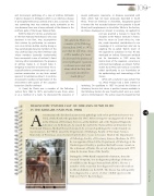
With the help of V. de Vos, a veterinarian of the then National Parks Board, who was stationed in the Park, they accomplished their mission by performing an examina- tion on an African buffalo (and by doing so they automatically became members of the very unofficial [but very elite!] Anthrax Club whose members [generally inadvertently] have necropsied a case of anthrax!). Apart from any other considerations, the presence of anthrax makes it, or should make it, obligatory to examine a blood smear micro- scopically before commencement of a post- mortem examination on any farm animal species if circumstances allow it. In so doing one prevents needless contamination of the environment with spores of the causative bacterium, Bacillus anthracis.
the disease in the
“J.L. (Jean) du Plessis was
a member of the Pathology Section from 1962 to 1975, and while he was there, alone or as a member of a team, he discovered the presence of several pathogenic organisms, or diseases associated with them, which had not been previously described in South Africa.”
and was granted a bursary to study the discipline at the Pasteur Institute in France. After his return to South Africa he, inter alia, made important contributions to our knowledge of E. ruminantium inter alia by adapting the so-called ‘Kümm strain’ of the organism to cultivation in mice. He also developed an indirect fluorescent antibody test for heartwater in 1981 using the Kümm stock of the organism, occurring in peritoneal macrophages, as antigen. Further studies by du Plessis and various co-workers added significantly to our knowledge on the epidemiology and immunology of the disease.
J.L. (Jean) du Plessis was a member of the Pathology Section from 1962 to 1975, and while he was there, alone or as a member of a team, he discovered the presence of
While still a student it was evident that J.G. (Pine) Pienaar had a keen interest in Pathology. On qualifying in 1957 he joined the Division of Veterinary Services but when a position became available in the Pathology Section he was ‘head-hunted’ and, as a result, came to Onderstepoort. The active research programme that
Aveterinarian who disclosed an interest in pathology early in his professional career was N.P.J. (Nick) Kriek who graduated in 1965. After serving for varying periods of time in the Division of Veterinary Services, at the Institute and at the Regional Diagnostic Laboratory in Stellenbosch, he joined the staff of the National Research Institute for Nutritional Diseases, Medical Research Council. As a research pathologist he worked mainly on mycotoxicoses. In 1980 he joined the Department of Pathology, Faculty of Veterinary Science, University of Pretoria as senior lecturer but left after 2 years to become Head of the Department
of Pathology, Faculty of Veterinary Science, Medunsa. In 1991 he was appointed Head of the Department of Pathology at the Onderstepoort Faculty, a position that had become vacant on the retirement of Tustin. He served in this position until 1999 when he became Dean of the Faculty.
During the 1990s Kriek’s main field of interest, from the research viewpoint, was in diagnostic pathology of diseases of wildlife in the Kruger National Park (KNP). He was a member of the team that first diagnosed bovine tuberculosis in African buffaloes in the KNP. The disease has subsequently been diagnosed in a variety of other wildlife species in the Park (see Part 3: Bacteriology).
147
147
Pathology
1908-2008
Years
